Publication Highlights
August 2026
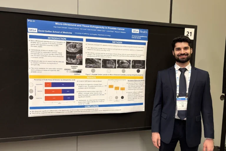
Micro-ultrasound tissue echogenicity predicts prostate cancer grade
This study explores how lesion echogenicity could be used to predict the presence of prostate cancer and its severity. Micro-ultrasound lacks a clear way of measuring tissue density, so the researchers tested whether echogenicity was a reliable sign of a patient's prostate cancer risk. The researchers found that hypoechoic lesions strongly indicated higher grade prostate cancers and lower ADC values than hyperechoic lesions. These findings suggest the consistency of echogenicity as an indicator of prostate cancer. This approach demonstrates that evaluating the shading of micro-ultrasound scans can significantly improve the accuracy of prostate scans. Overall, doctors will be able to make better decisions on patient treatment and avoid unnecessary biopsies. To access the full article, click the link below.
A Histopathologic Assessment of Prostate Ductal Anatomy in Relation to Micro-ultrasound
This study investigated how the ductal structures of the prostate may influence the appearance of cancer on micro-ultrasound imaging. By analyzing prostate tissue samples after biopsy, researchers evaluated ductal features within benign and cancerous regions to determine whether these characteristics could explain differences observed on imaging. The findings suggest that variations in prostate ductal structure may serve as a potential imaging biomarker, providing new insight into how micro-ultrasound detects and characterizes prostate cancer. This approach may contribute to improved visualization of clinically significant disease and support more accurate, targeted diagnosis. To access the full article, click the link below.
Detection Rate of Prostate Cancer Using Prostate-Specific Membrane Antigen/Ultrasound-Fusion Biopsy in Patients with Suspected Recurrence or Inconclusive/Negative Biopsies: A Single-Center Retrospective Study
This study discusses the diagnostic challenge of detecting prostate cancer in patients with suspected recurrence after prior treatment or with previous inconclusive/negative biopsies. To improve detection, researchers at UCLA combined PSMA PET and CT imaging with ultrasound-fusion biopsy, allowing biopsies to be precisely guided toward PSMA-avid lesions rather than relying on standard sampling alone. Cancerous lesions consistently showed higher PSMA uptake (SUVmax), larger size, and higher visual PRIMARY scores than non-cancerous lesions, with detection rates climbing sharply as PRIMARY scores increased. Detection rates were slightly lower in patients previously treated with non-surgical therapies compared to treatment-naïve patients, suggesting prior treatment may alter PSMA uptake patterns. Because this approach integrates molecular imaging with precise biopsy targeting, it offers improved accuracy for identifying clinically significant cancer in difficult-to-diagnose patients. This precision may help urologists better target biopsies, reduce missed diagnoses, and tailor follow-up strategies based on both imaging findings and individual patient treatment history. To access the full article, click the link below.
Head-to-head Comparison of Multimodal Imaging for Prostate Cancer Detection and Local Staging with Whole-mount Histopathology Reference
Accurate localization and staging of clinically significant prostate cancer are critical for guiding biopsy, treatment selection, and surgical planning. While multiparametric MRI (mpMRI) has become the standard imaging modality for prostate cancer evaluation, emerging technologies such as micro-ultrasound (MicroUS) and prostate-specific membrane antigen positron emission tomography/computed tomography (PSMA PET/CT) have shown promise in improving cancer detection and characterization. However, direct comparisons between these imaging modalities using whole-mount histopathology—the gold standard for assessing tumor location and extent—have remained limited.This was the first study to compare the diagnostic performance of micro-ultrasound (MicroUS), multiparametric magnetic resonance imaging (mpMRI), and prostate-specific membrane antigen positron emission tomography/computed tomography (PSMA PET/CT) for detecting and locally staging prostate cancer, using whole-mount histopathology from radical prostatectomy specimens as the reference standard. To access the full article, click the link below.