Projects & Publications
Featured Publications
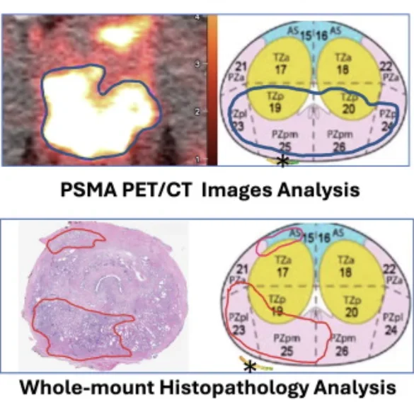
Detection Rate of Prostate Cancer Using Prostate-Specific Membrane Antigen/Ultrasound-Fusion Biopsy in Patients with Suspected Recurrence or Inconclusive/Negative Biopsies: A Single-Center Retrospective Study
This study discusses the diagnostic challenge of detecting prostate cancer in patients with suspected recurrence after prior treatment or with previous inconclusive/negative biopsies. To improve detection, researchers at UCLA combined PSMA PET and CT imaging with ultrasound-fusion biopsy, allowing biopsies to be precisely guided toward PSMA-avid lesions rather than relying on standard sampling alone. Analyzing 62 patients and 89 lesions, the researchers found that this fusion approach detected cancer in 66% of patients and 58% of lesions overall. Cancerous lesions consistently showed higher PSMA uptake (SUVmax), larger size, and higher visual PRIMARY scores than non-cancerous lesions, with detection rates climbing sharply as PRIMARY scores increased. An SUVmax threshold of 7.07 was identified as the optimal cutoff for distinguishing cancerous lesions from benign lesions. Detection rates were slightly lower in patients previously treated with non-surgical therapies compared to treatment-naïve patients, suggesting prior treatment may alter PSMA uptake patterns. Because this approach integrates molecular imaging with precise biopsy targeting, it offers improved accuracy for identifying clinically significant cancer in difficult-to-diagnose patients. This precision may help urologists better target biopsies, reduce missed diagnoses, and tailor follow-up strategies based on both imaging findings and individual patient treatment history.
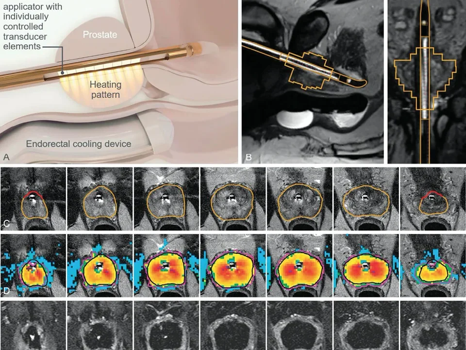
Perioperative outcomes from a randomized controlled trial comparing MRI-guided transurethral ultrasound ablation (TULSA) to robot-assisted radical prostatectomy (RARP)
This study evaluates how MRI-guided transurethral ultrasound ablation (TULSA) can improve tissue ablation results. The less-invasive TULSA outcomes were compared to those of robot-assisted radical prostatectomy. (RARP) The researchers found that patients under TULSA experienced lower blood loss, shorter hospital stays, and decreased pain in comparison to RARP. Although TULSA patients faced a longer recovery time, their recovery was overall more successful, resulting in better health outcomes. This method shows how ultrasonic ablation can be less taxing on patients and offering a better recovery experience. Doctors may be able to provide this less intrusive option for medium grade prostate cancers while still yield greater results than RARP.
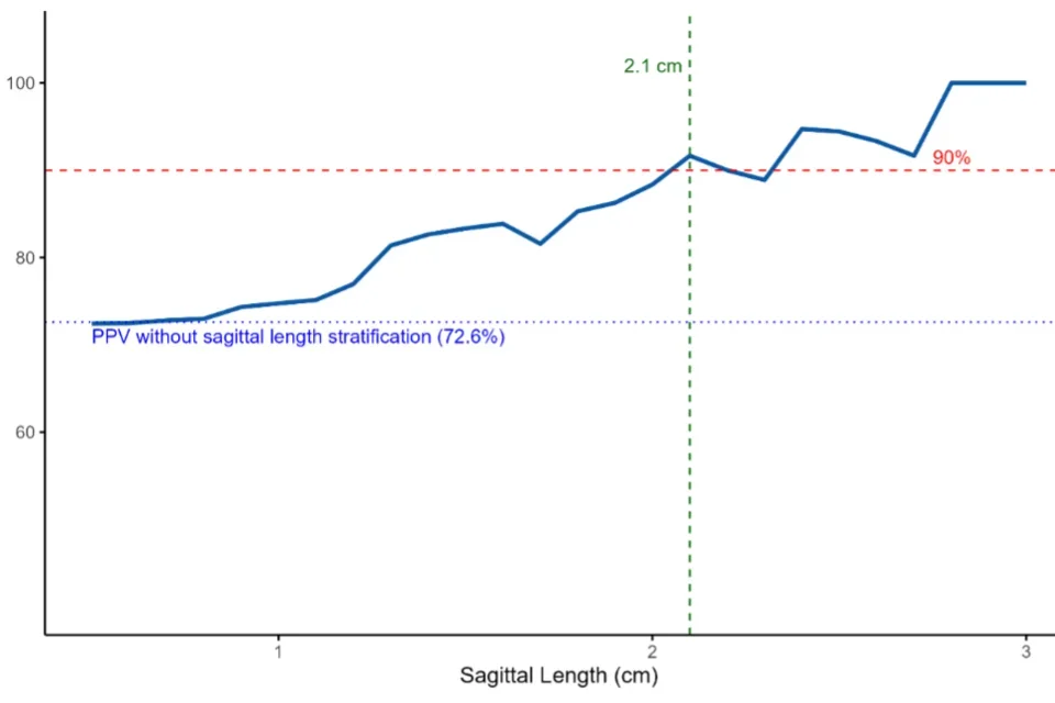
Improving prostate cancer detection using sagittal length on micro-ultrasound
Detecting prostate cancer has always been a challenge in the field of urology. One new imaging tool, micro-ultrasound, has shown to detect prostate cancer in ways that differ from conventional MRI. The authors’ study investigates whether the size of a suspicious cancerous area on micro-ultrasound can improve the prediction of prostate cancer in areas seen on both MRI and micro-ultrasound (dual-image visible lesions). Previously, the group found that dual-image visible lesions predicted prostate cancer 72% of the time. In this study, they found that those same areas with a length of 2.1 cm or more on micro-ultrasound increases the prediction rate to 90%. This finding provides urologists and patients with more information about the odds of prostate cancer and allows the patient to make an informed at the time of biopsy.
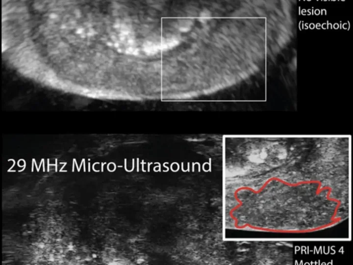
Combining Prostate MRI and Microultrasound to Omit Systematic Biopsy
Prostate cancer diagnosis involves biopsy. MRI guided biopsy occasionally misses prostate cancer. Systematic template biopsies are added in order to detect missed cancer. However, these biopsies frequently return without finding cancer and increase patient related side effects. This study explored combining MRI and high resolution micro-ultrasound to visualize the target using two separate imaging modalities and thus omit systematic biopsies. The dual imaging technique identified 97.4% of clinically significant prostate cancer while systematic biopsy alone detected 61.5%. These findings suggest that combining MRI and micro-ultrasound may help doctors more accurately target cancer and potentially reduce or omit systematic biopsies in select patients.
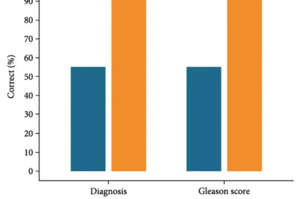
Patient Educational Videos Delivered With Prostate Pathology Results Improve Knowledge and Satisfaction
This study looks at how short educational videos can help patients better understand their prostate biopsy results. Medical information can be confusing, especially during stressful moments, so the researchers tested whether simple videos could make a difference. They found that patients who watched the videos had a better understanding of their diagnosis and felt more satisfied with their care. The videos helped explain complex terms in a clear and easy way. This approach shows how better communication tools can improve the patient experience. By helping patients feel more informed and confident, these videos may support better decision-making and stronger relationships between patients and their care teams.
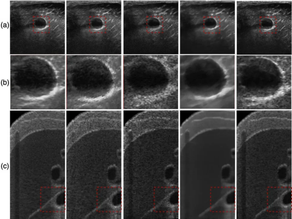
Geometry-Aware Implicit Neural Reconstruction of Oblique Micro-Ultrasound Scans
This study addresses issues of micro-ultrasound scans producing unaligned oblique slices with standard MRI or histopathology. To combat this, researchers created a geometry-based framework that reshapes oblique slices into sharp, axial 3D volumes. The researchers found that the new model was able to produce clearer 3D imaging in comparison to a traditional interpolation model. The texture and boundaries of the ultrasound are preserved and help crucial identification of cancer. Because this approach has improved adjustments for processing angled ultrasound data, higher accuracy of diagnosis can be achieved. This precision will allow doctors to have smoother and faster evaluations, as they can reliably monitor tumors and identify prostate cancer.
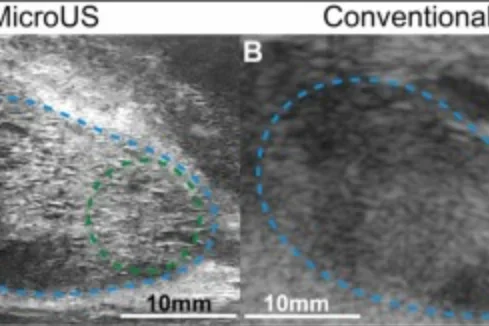
A Histopathologic Assessment of Prostate Ductal Anatomy in Relation to Micro-ultrasound
This study investigated how the ductal structures of the prostate may influence the appearance of cancer on micro-ultrasound imaging. By analyzing prostate tissue samples after biopsy, researchers evaluated ductal features within benign and cancerous regions to determine whether these characteristics could explain differences observed on imaging. The findings suggest that variations in prostate ductal structure may serve as a potential imaging biomarker, providing new insight into how micro-ultrasound detects and characterizes prostate cancer. This approach may contribute to improved visualization of clinically significant disease and support more accurate, targeted diagnosis.
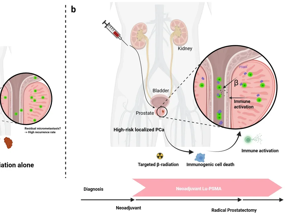
177Lu-Prostate-Specific Membrane Antigen Neoadjuvant to Stereotactic Ablative Radiotherapy for Oligorecurrent Prostate Cancer (LUNAR): An Open-Label, Randomized, Controlled, Phase II Study
This study explores the effects of targeted prostate-specific membrane antigens (PSMA) on stereotactic body radiotherapy (SBRT). Because radiation can miss microscopic cancer cells, the researchers tested whether adding neoadjuvant 177Lu-PNT2002 could safely destroy these invisible spots. The reserachers found that this radioligand therapy was able to significantly extend progression-free survival from 7.4 months to 17.6 months. These findings demonstrate a hazard ratio of 0.37, representing a 63% reduction in the risk of progresion, salvage hormonal therapy, or death without an attendant increase in toxicity. This approach demonstrates that using targeted radioligand therapy can significantly improve eugonadal progression-free survival. Overall, clinicians will be able to optimize oncology outcomes and succesfully prolong therapy-free survival for patients.
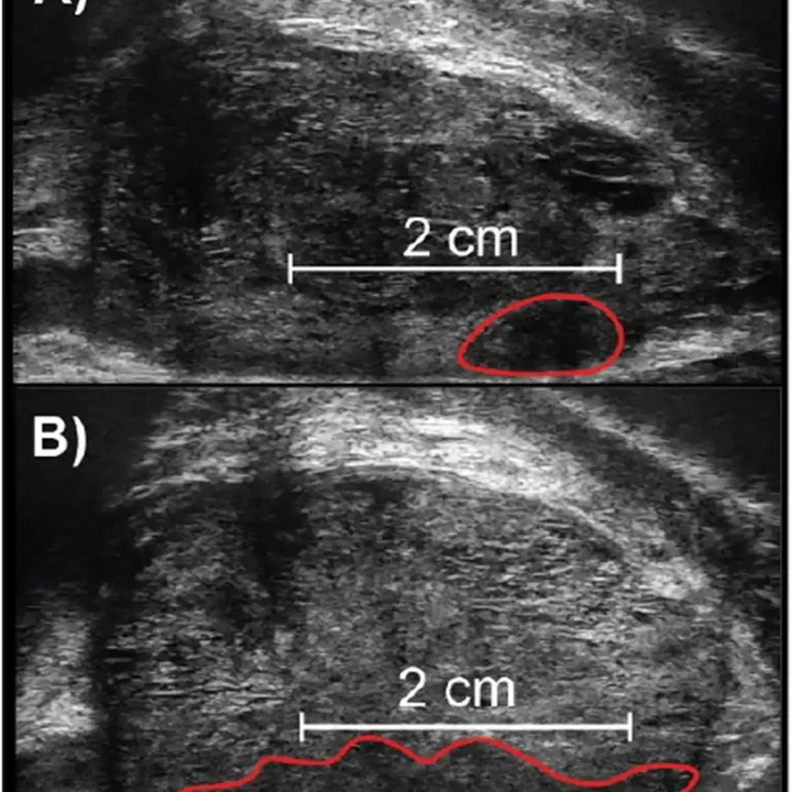
Using Sagittal Length to Differentiate Between PRI-MUS 4 and PRI-MUS 5 Lesions
This study looks at whether measuring lesion size can improve how doctors classify prostate cancer risk using micro-ultrasound imaging. Currently, the PRI-MUS grading system relies only on how a lesion looks (its acoustic pattern) to separate high-risk (score 4) from very-high-risk (score 5) lesions, unlike MRI's PI-RADS system, which also factors in lesion size. The researchers analyzed 275 PRI-MUS 4 and 5 lesions from patients who underwent micro-ultrasound-guided biopsy at UCLA. They tested whether adding a size measurement, called sagittal length, could better predict which lesions actually contained clinically significant cancer. They found that lesions of 2 cm or larger were meaningfully more likely to be cancerous. Using this 2 cm cutoff to reclassify lesions improved the cancer-detection accuracy of PRI-MUS 4 lesions by about 10%, without reducing the accuracy of PRI-MUS 5 lesions.
Because this approach uses a simple, objective measurement rather than subjective visual interpretation, it could make micro-ultrasound grading more consistent between different doctors and clinics. Overall, this technique could help reduce inconsistency in prostate cancer risk assessment, guide more precise biopsy decisions, and bring micro-ultrasound classification closer in line with standardized MRI-based approaches.
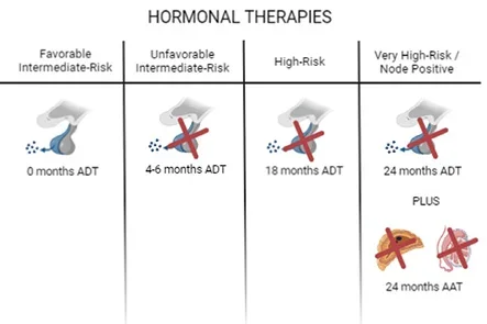
Hormone therapy use and duration with postoperative radiotherapy for recurrent prostate cancer: an individual patient data meta-analysis
After surgery to remove the prostate, cancer can come back, resulting in doctors treating the remaining cancer with radiation. This study asked whether adding hormone therapy to radiation helps with patient survivorship and if the duration of hormone therapy can change patient outcomes. Researchers pooled data from over 6,000 patients across six major randomized clinical trials to perform an analysis. Overall, they discovered the addition of hormone therapy did not significantly improve survivorship, especially for men whose PSA (a known marker of cancer activity) was low at the time of radiation. However, men with higher PSA levels before treatment showed significant responses to hormone therapy treatment, as hormone therapy offered meaningful survival benefit. Short-term hormone therapy worked as well as long-term therapy within the group. These findings suggest hormone therapy as a treatment should not be routinely added to radiation for most men in early stages of treatment, but it may be worthwhile for those with higher PSA levels.
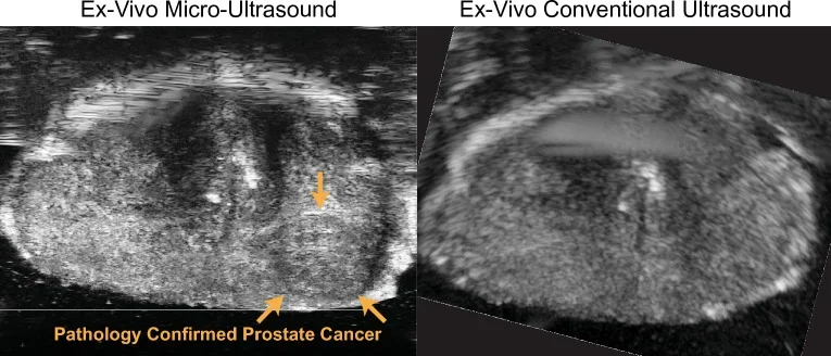
Evaluation of Prostate Cancer Detection Using Micro-Ultrasound Versus MRI through Co-Registration to Whole-Mount Pathology
Micro-ultrasound has recently been introduced as a low-cost alternative to multi-parametric MRI for imaging prostate cancer. Early clinical studies have demonstrated promising results; however, robust validation via comparison with whole-mount pathology has yet to be achieved. Due to micro-ultrasound probe design and tissue deformation during scanning, it is difficult to accurately correlate micro-ultrasound imaging planes with ground truth whole-mount pathology slides. In this study, we developed a multi-step methodology to co-register micro-ultrasound and MRI to whole-mount pathology.
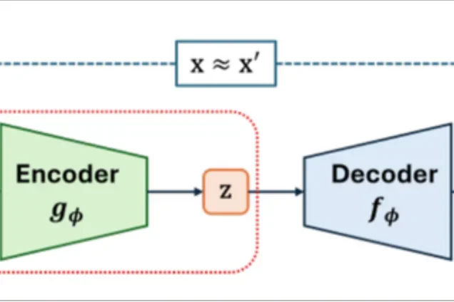
AI-Enhanced Micro-Ultrasound Improves Detection of Clinically Significant Prostate Cancer at Biopsy
This study explores a new way to find important prostate cancer using artificial intelligence (AI) and micro-ultrasound imaging. Micro-ultrasound provides very detailed images of the prostate, and AI helps highlight areas that may be cancer. The researchers found that this method can improve how doctors detect serious cancers during a biopsy. It may also reduce unnecessary biopsies by helping doctors focus only on the most suspicious areas. Because this approach can be done in real time during a clinic visit, it may offer a faster and more accessible option than traditional imaging methods like MRI. Overall, this technique could help doctors diagnose prostate cancer earlier and more accurately, leading to better patient care.

The Relationship Between Cannabis and Male Infertility, Sexual Health, and Neoplasm: A Systematic Review
In the United States of America (USA), cannabis is legal in 28 states for medical purposes and 8 states for recreational use. In 2016, the legal marijuana industry reached nearly $7 billion in sales in the USA alone. Although consumption continues to increase, the medical effects of marijuana remain understudied. Young males comprise the demographic most likely to consume cannabis, and these individuals will be most vulnerable to its short- and long-term consequences. The purpose of this manuscript is to systematically review the available literature describing the effects of marijuana on male infertility, sexual health, and urologic neoplasms.
Read More About Cannabis Impacts on Male Fertility